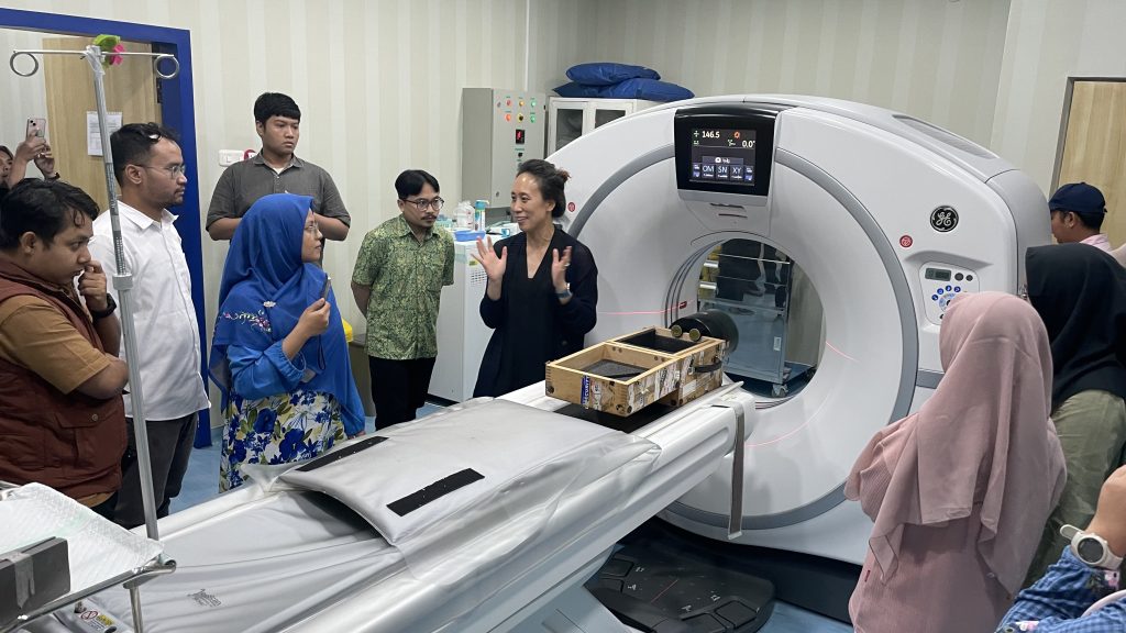
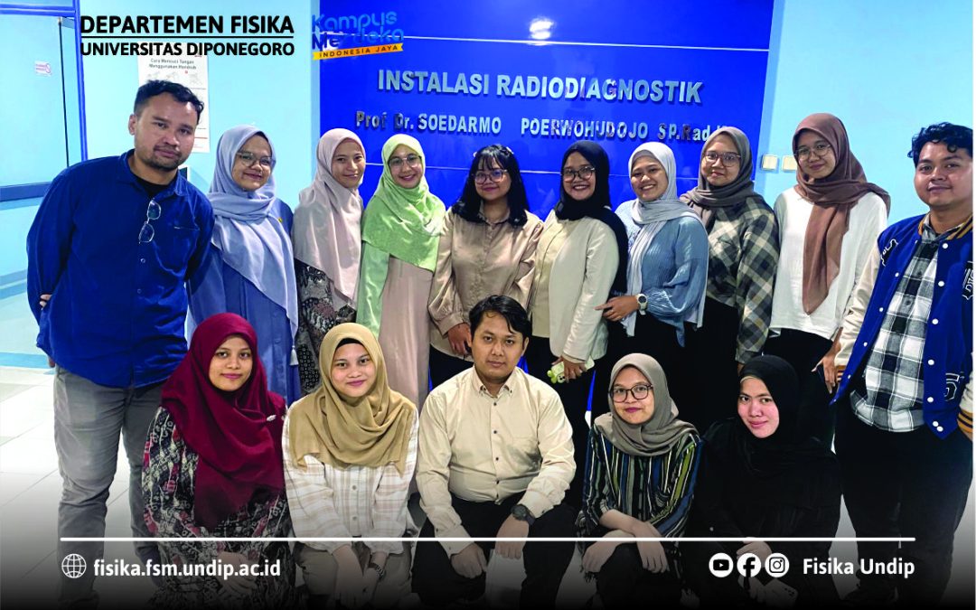
The Institute of Electrical and Electronics Engineers – Nuclear and Plasma Sciences Society (IEEE NPSS) has organized a summer school entitled School on Advanced Topics in Medical Imaging. The school was held for 5 days, from July 31 – August 4, 2024 at the University of Indonesia and Dharmais Cancer Hospital. Registration as a student has been done through an online selection system. A total of 15 Diponegoro University Master of Physics students were selected to attend this school from a total of 28 students.

The main goals of the school are to train master level students in the area of hybrid PET/CT and PET/MR imaging, to encourage the participation of young scientists in the field of medical imaging, and to provide a refresher and enrichment of knowledge on PET/CT and PET/MR for medical physicists working with these technologies. These goals are achieved through a combination of classroom lectures and exercises in a minilab and hospitals. According to the information on the school’s official website (https://indico.cern.ch/event/1353971), specific topics are delivered on an organized schedule as shown in the timetable below.

This school is a collaborative activity between IEEE NPSS and the UI Physics Department, and is fully funded by IEEE. The faculty members in this school come from various countries and expertise, namely Dr. Martin Grossmann (Paul Scherrer Institute, Switzerland) as coordinator, Dr. Andrea Gonzales-Montoro (Universitat Politecnica de Valencia, Spain), Dr. Kaitlin Hellier (University of California, Santa Cruz, USA), Prof. Steven Meikle (University of Sydney, Australia), Prof. Yungho Seo (University of California, San Francisco, USA), and Dr. Zhye Yin (GE Healthcare).
Medical imaging is a great method for diagnosing diseases. In patients suspected of having cancer, imaging using only CT may not be sufficient to conclude the diagnosis. CT images provide excellent detail for anatomical information, but are insufficient to provide metabolic and functional information. On the other hand, imaging using PET is excellent for generating functional information in the form of abnormal activities that may occur in patients with cancer cells. However, PET images do not produce detailed information about the patient’s anatomy. By combining the advantages of both technology (PET/CT), the resulting hybrid image will be able to help identify radioactivity in the organs inside the patient.
From this school, the students gained a lot of insight into the fundamental basis of medical imaging, image reconstruction and registration, radiopharmaceutical production, types and functions of various X-ray detectors, dose evaluation and image quality, etc. A number of practicum sessions were also conducted, namely EasyPET in the minilab, CT, planar, and PET/CT imaging in hospitals. In addition, students also gained networking experience with the lecturers to continue learning and conducting research related to similar topics.


